Multiparametric Ultrasound Assessment of Alopecia Areata Plaque with Atrophy After Serial Corticosteroid Infusion
DOI:
https://doi.org/10.62742/2965-7911.2025.2.bjhh24Keywords:
Alopecia Areata, Multiparametric Ultrasound, Shear Wave Elastography, Corticosteroid InfusionAbstract
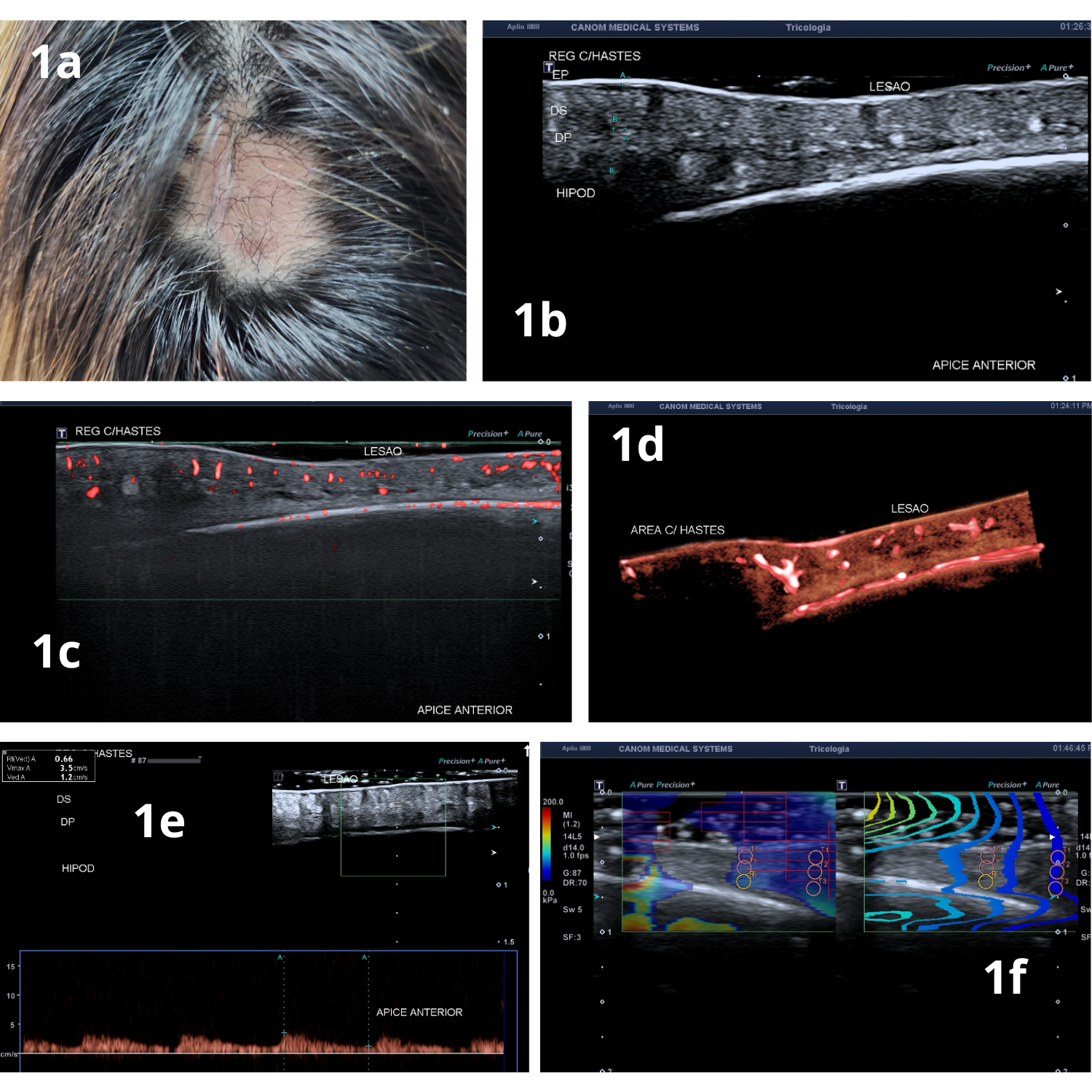
Introduction: This study explores the use of multiparametric ultrasound (MPUS) to evaluate a case of alopecia areata (AA) with scalp atrophy resulting from repeated corticosteroid injections. AA is a non-scarring, autoimmune alopecia characterized by lymphocytic infiltration near the hair follicle bulb, causing hair loss. Excessive corticosteroid use can lead to tissue atrophy and impaired hair growth. The patient, a 27-year-old woman, presented with a resistant scalp lesion following multiple corticosteroid injections.
Perspectives: The MPUS examination combined B-mode imaging, Doppler ultrasound, and shear wave elastography. B-mode imaging revealed scalp thinning (2.4 mm vs. 4.9-5.0 mm in adjacent areas), while Doppler showed reduced vascular density and tortuous vessels with a peak systolic velocity of 3.5 cm/s (normal: 5.5 cm/s). Elastography indicated increased tissue stiffness in the reticular dermis and hypodermis compared to surrounding areas. The study concludes that MPUS is an effective non-invasive diagnostic tool, providing anatomical and functional insights into scalp conditions, thus offering an alternative to biopsy for monitoring therapeutic outcomes.
Downloads
Downloads
Published
Issue
Section
License
Copyright (c) 2025 Brazilian Journal of Hair Health

This work is licensed under a Creative Commons Attribution 4.0 International License.


